Navicular syndrome in horses
The most important facts at a glance
What is the navicular syndrome?
- The hoof is a complex shock absorber apparatus in the lower part of the horse's leg. If it is damaged, this is known as navicular syndrome or podotrochlosis.
- Typical symptoms include lameness, stumbling, a stiff gait or a restrictive posture.
- The main causes are genetic predisposition, lack of exercise, incorrect shoeing and overloading.
- Timely diagnosis and treatment increase the chances of the horse remaining rideable.
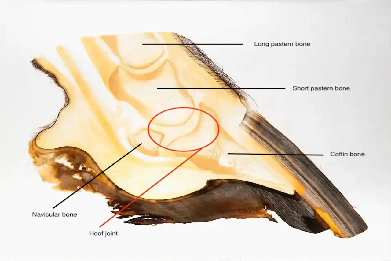
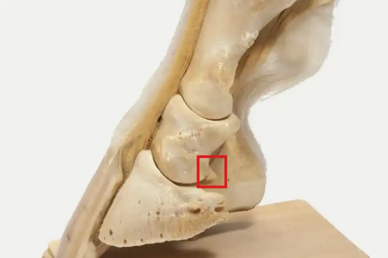
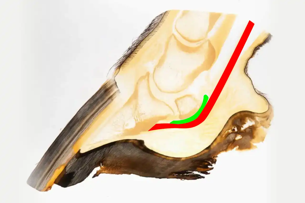
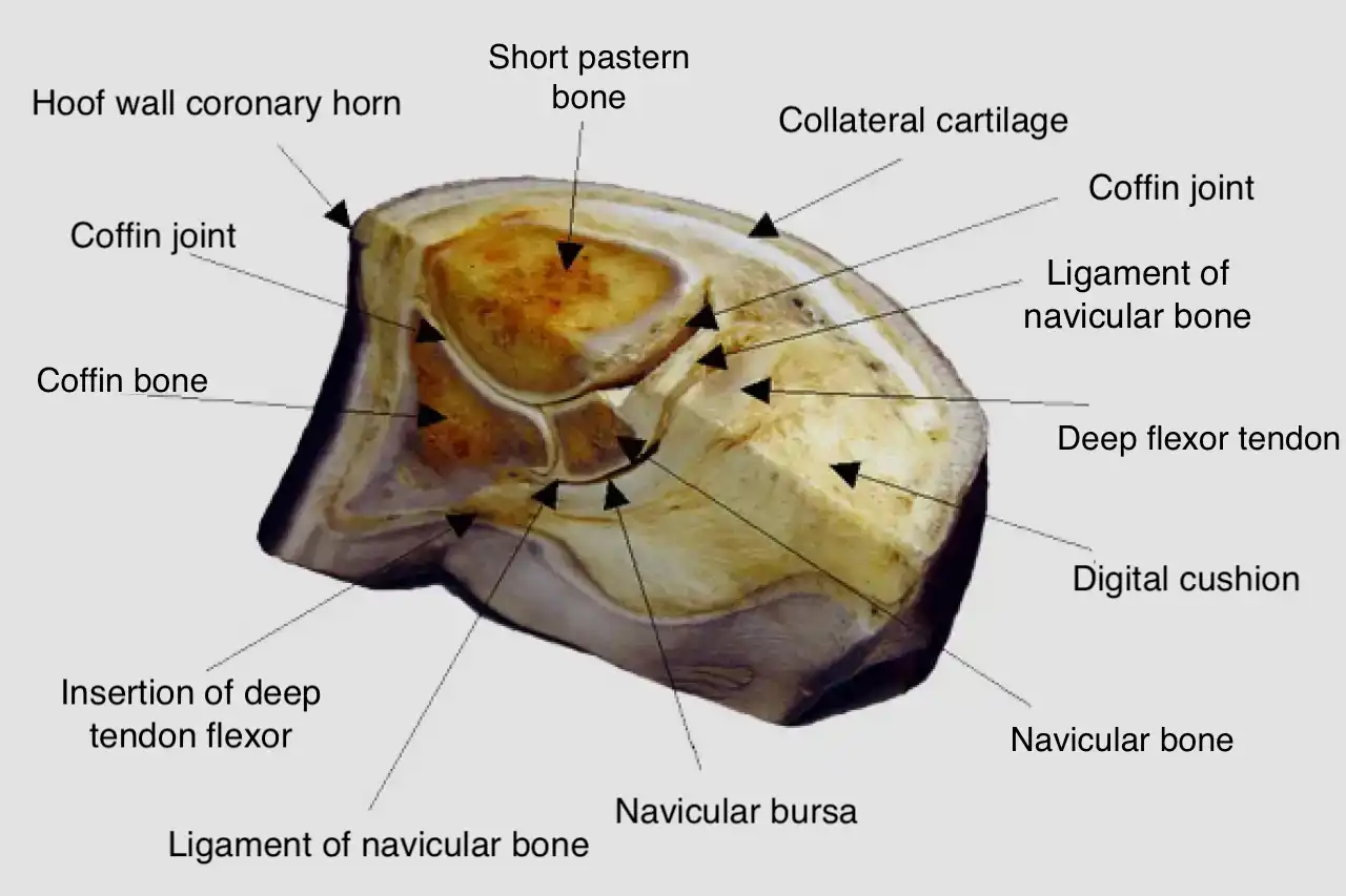
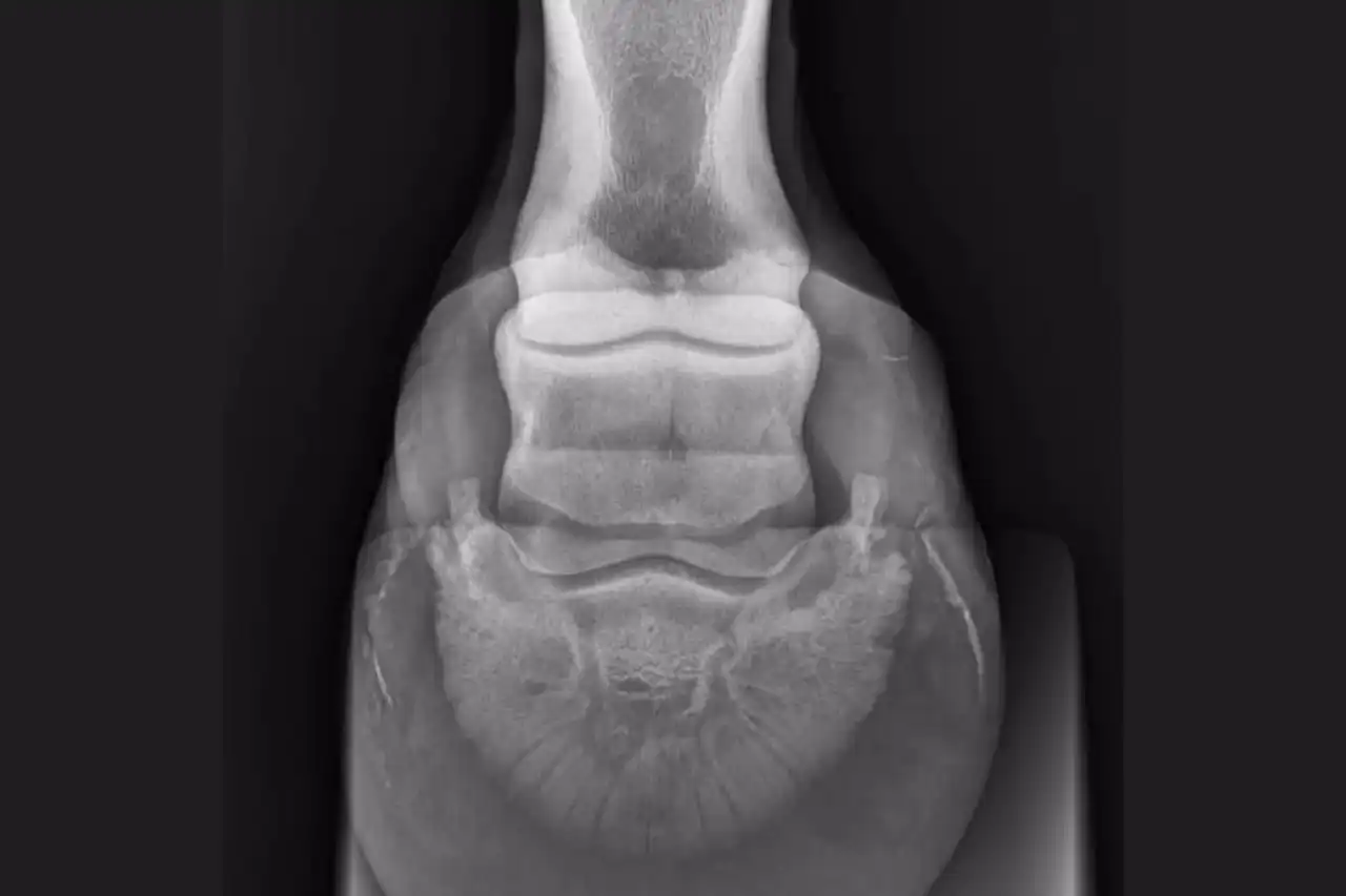
Three bones meet in the hoof or distal phalangeal joint (medical term: articulatio interphalangealis distalis), i.e. the joint between the last two phalanges: the coffin bone (distal), the short pastern bone (middle) and the navicular bone (also known as the sesamoid bone). The navicular bone (Latin: Os sesamoideum distale) is a small sesamoid bone that is formed on the deep flexor tendon in hoofed animals. There is one navicular bone per toe. It is located below the tendon in the area of the joint between the coffin bone and the short pastern bone. Between the deep flexor tendon and the navicular bone lies the bursa podotrochlearis, which serves as a gliding bearing. The navicular bone, bursa and deep flexor tendon together form the podotrochlear region.
The articular cartilage of this hoof joint is comparatively thick and serves as a shock absorber. The joint has to withstand heavy loads, especially when walking, as it has to continuously compensate for uneven ground.
Anatomy and function of the podotrochlear region
The podotrochlear region is made up of several structures:
- Coffin bone
- Flexor tendon
- Lateral ligament
- Navicular bursa
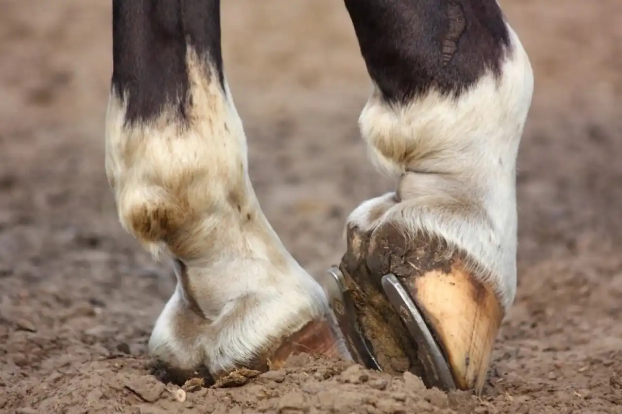
This region acts as a shock absorber with every step and distributes the forces evenly over the hoof and leg. In the event of permanent overloading or inflammation, the navicular bone can degenerate or even break. The disease almost exclusively affects the front hooves and is often confused with laminitis. The chronic form is known as podotrochleosis.
Symptoms and diagnosis
What are the first signs of the navicular syndrome?
- Stiff or lame gait, especially after periods of rest
- Stumbling and fidgeting at the start of movement
- Careful posture, reduced enjoyment of movement
How is the navicular syndrome diagnosed?
- Clinical examination by the vet
- R&R images are often unspecific
- Exact localization by nerve blockade: If the horse is lame-free after a targeted anaesthetic, the origin lies in the podotrochlear region.
Causes of the navicular syndrome
Navicular syndrome occurs exclusively in domesticated horses. The causes are manifold:
- Genetics: predisposition through breeding lines
- Improper loading: due to hard stops, jumping, tight turns in equestrian sports
- Lack of exercise: especially harmful at a young age
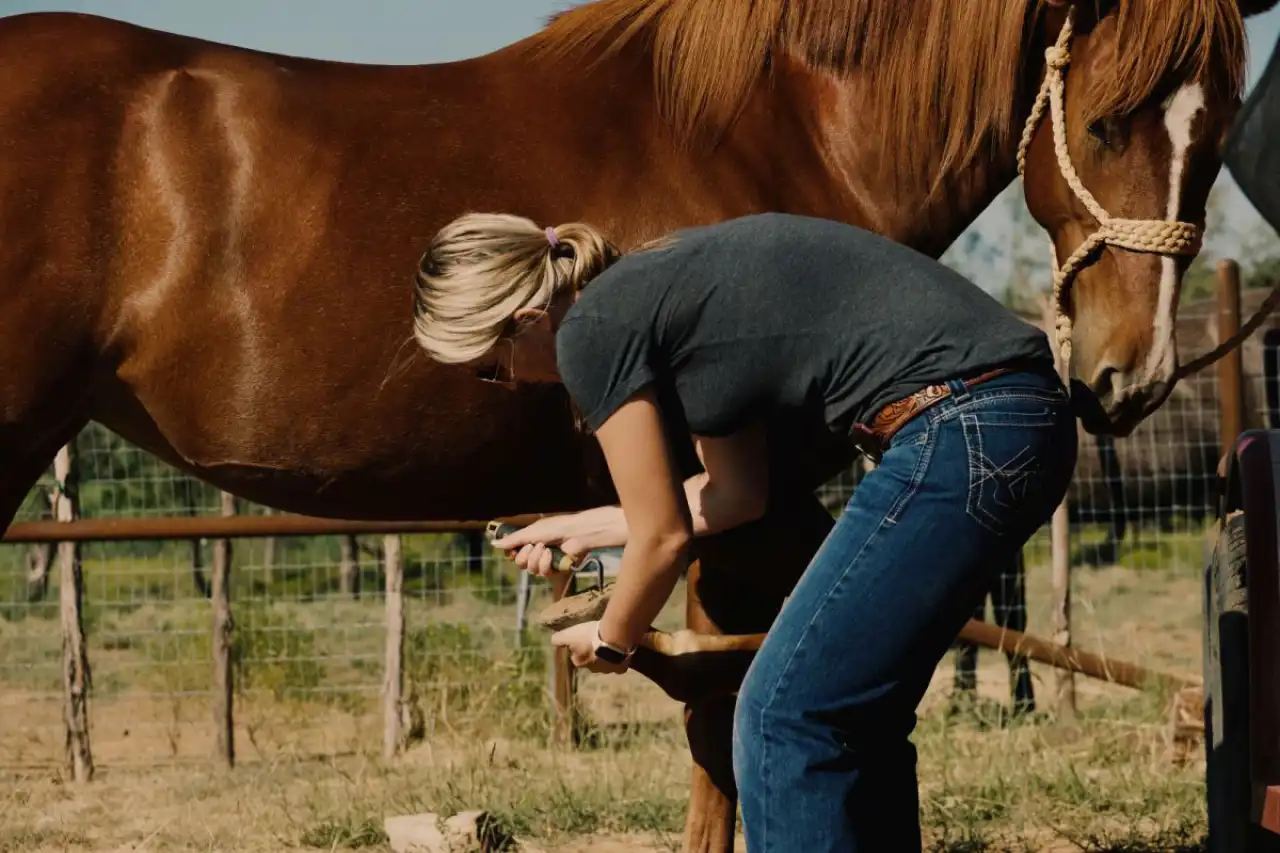
- Misalignment and hoof trimming: toes that are too long, incorrect shoeing or horseshoes that are too tight
Treatment and therapy
What helps?
- Rest phase for acute inflammation
- Anti-inflammatory medication
- Targeted exercise on soft ground
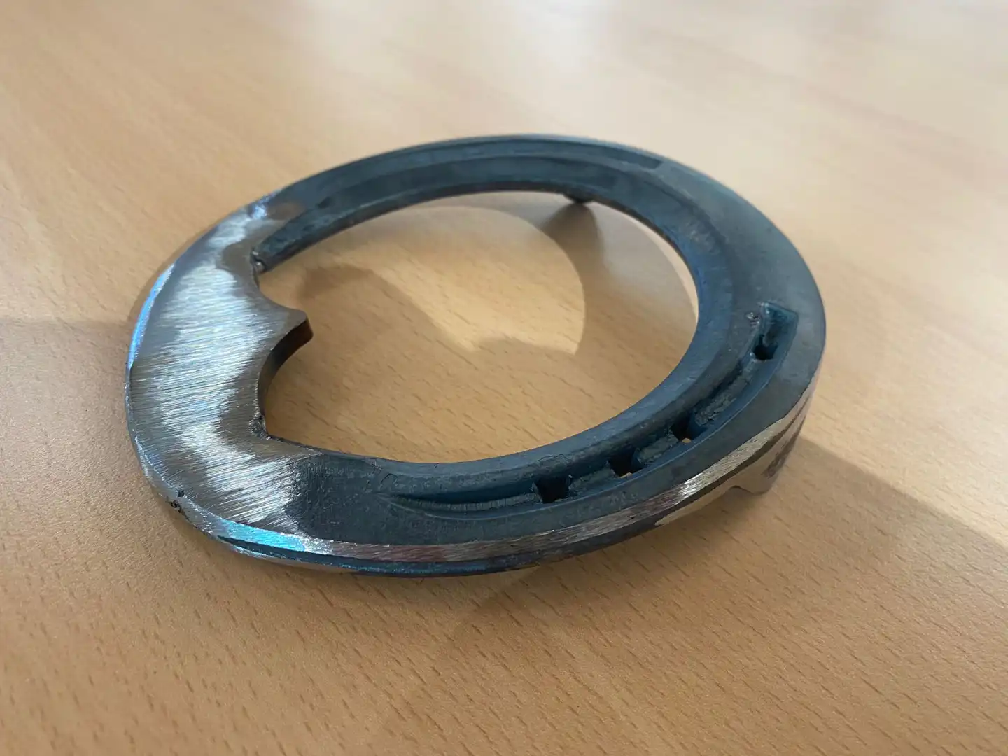
- Orthopaedic shoe for relief *
- Ground work as a gentle training method
Alternative remedies such as devil's claw or homeopathic preparations can be used as a supportive measure, but should be agreed with the vet.
* Rounding off the toe to improve movement is recommended here. The rolling of the hoof, i.e. its rotational movements in all directions, should thus be facilitated. Welding a steel-aluminum piece between the ends of the rod ends or a bar shoe can be used for support. Wedge pads (preferably made of leather or combi-leather-plastic) can also provide relief.
What is no longer recommended?
- A nerve incision to suppress lameness is no longer appropriate and carries considerable risks (risk of tripping, accidents).
Forecast
Does my horse need to be put down?
In most cases, no. If the disease is recognized early and treated correctly, the horse can be ridden again. Only in the case of advanced, painful destruction of the structures must euthanasia be considered - but this is the exception.

